Phone Number:0755-26943630

News Center/ 新闻中心

Medical endoscope industry research report

Time:2020-07-25 23:34 Click:/Second

1. Definition
 
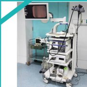
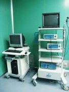
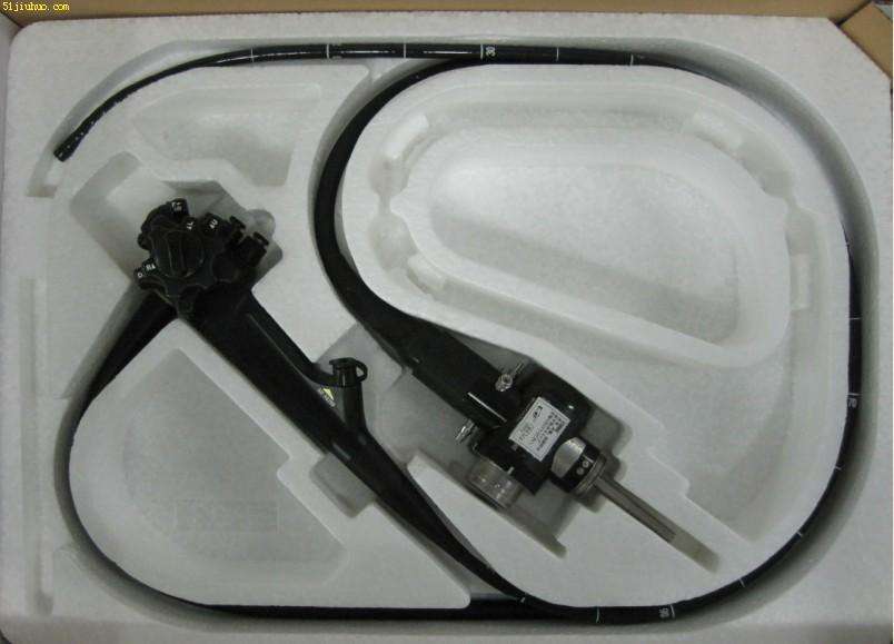
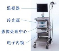
The endoscope is composed of three parts: a speculum system, an imaging system and a lighting system. It is a collection of optics, ergonomics,
 
Precision machinery, modern electronics, mathematics, IT and other technical testing instruments. Endoscopes are divided into industrial endoscopes and medical endoscopes.
 
A medical endoscope is an invasive inspection tool that enters the human body through a natural orifice of the human body or a small incision made by surgery, which can help doctors observe the pathological changes of human organs that cannot be shown by X-rays. In clinical practice, it enters the human body through the natural orifice or percutaneous puncture channel of the human body, and is introduced into the organ to be examined or operated for optical imaging, so as to provide the doctor with image information for disease diagnosis, and the operation can be performed with the cooperation of equipment treatment. The medical endoscope is one of the medical devices used by medical personnel to observe the diseased tissue inside the human body directly and effectively. It has the advantages of high image clarity and simple operation. With the development of technology, the scope of use of endoscopes is gradually expanding, and it is more closely integrated with treatment, and the frequency of use in clinical diagnosis and treatment is increasing. At present, endoscopes have been widely cited in the fields of otolaryngology, abdominal surgery, urology, anorectal surgery, orthopedics, thoracic and cardiovascular surgery, neurosurgery, and gynecology.
 
2. The development process and principle of technology
 
The English name of endoscope is endoscopy, which originated from Greek, and the Latin letter "endo" means "inside". About AD
 
Between the first 460 and the first 370, the Greek physician Hippocrates once described a rectal examination device, which uses natural light sources to examine the rectum, cervix and other parts of the patient. Hippocrates is known as the "originator of endoscopy."
 
The real development of the endoscope system originated in modern times, with a development process of more than 200 years, from the initial rigid tube endoscope (1806), semi-flexed endoscope (1932) to fiber endoscope (1957), Then to today's electronic endoscope (1983). The image quality of medical endoscopes continues to improve, and its clinical applications are becoming more and more popular.
 
(1) Rigid tube endoscope (1806-1932):
 
In 1804, German Philip Bozzini (Philip Bozzini) proposed the idea of ​​an endoscope, and in 1806 completed a candle as a light source, consisting of a vase-like light source, a candle and a series of lenses The composition used to observe the internal structure of the bladder and rectum of animals is called the Lichtleiter (Light Conductor), so Philip is the inventor of modern endoscopes.
 
In 1853, French doctor De Solmo invented the genitourinary endoscope, so he is known as the "father of endoscope"; in 1879, the German urologist Nitze (Nitze) and the Vienna equipment manufacturer Leiter made it. In addition, the first endoscope with an optical system (ie, cystoscope) was introduced, which used circulating ice water to avoid damage, and obtained clearer images and gradually introduced the operation pipeline. In 1881, Wright, who parted ways with Dr. Nietzsche, and German doctor Mikulicz used Nietzsche's rigid tube optical system to successfully make the first clinical gastroscope. In 1898, the German laryngologist Kirian ( Killian) made and successfully used the first bronchoscope.
 
(2) Semi-flexible endoscope (1932-1957):
 
It was developed by German doctor Rudolf Schindler in cooperation with equipment maker John Wolf (Georg. Wolf) in 1928, and finally succeeded in 1932, named Wolf-Schindler The birth of gastroscope means the development of semi-flexible endoscope. Its characteristic is the flexibility of the front end, that is, there is a certain range of bending in the stomach, so that the surgeon can clearly observe the image of the gastric mucosa. The gastroscope has a smooth metal ball on the front end, which is convenient to insert, the light bulb is bright, and there is an air channel. Used to inject air, the proximal end is a hard tube, with eyepieces for focusing. This gastroscope has become the standard product in the gastroscope field for more than two decades.
 
Dr. Odolph Schindler has not only made great achievements in the development and transformation of gastroscopy, he is also one of the most active gastroscopy promoters in history. He published "Gastroscopy Textbook and Atlas" ("Lehrbuch und Atlas der Gastroskopie") reported more than 400 cases of uncomplicated gastroscopy for the first time, making an outstanding contribution to the promotion and application of gastroscopy. One defect of Schindler's gastroscope products is that the lighting system uses the heat source of incandescent lamps, which has certain risks, such as possible burns in the body cavity and accidents caused by electrical failure of the incandescent lamp.
 
 In 1952, the French team of scientists Fourestier, Gladu and Valmiere produced a cold light source glass fiber lighting device, which greatly improved the safety of laparoscopy and laid the foundation for the subsequent application of flexible fiber instruments.
 
(3) Fiber endoscope (after 1957):
 
In 1954, based on the theoretical basis that the fiber conductive light source and the inter-fiber coating can solve the optical insulation between the fibers, the British Hopkings and Kapany studied the precise arrangement of the fibers, which effectively solved the image of the fiber bundle. Transmission has laid the foundation for the practicality of fiber optics.
 
In 1957, South African doctor Hirschowitz and two physicists made the world's first prototype of an optical fiber endoscope for examining the stomach and duodenum. In 1960, American Cystoscopy Manufacturers (ACMI) provided Hirschowitz with the first commercial fiber endoscope. In the same year, Karl Storz, the founder of Storz, and his team developed Cold light. In 1966, Karl Storz and Hopkins produced a cylindrical lens system. In 1967, Machida company adopted an external cold light source, which greatly increased the brightness of the light, and found small lesions and further expanded the field of view. With the continuous improvement of accessory devices, such as the development of surgical instruments and photography systems, fiber endoscopes can be used not only for diagnosis but also for surgical treatment.
 
(4) Electronic endoscope (after 1983):
 
In 1983, the Welch Allyn Company of the United States developed and applied high-performance micro image sensors (charge coupled device, CCD) to replace the fiber optic imaging technology of endoscopes, announcing the birth of electronic endoscopes, which is considered to be another time in the history of endoscopes. Historic breakthrough. After 2000, the development of ultrasound probes and three-dimensional probes, and the advent of new electronic endoscopes such as capsule gastroscopes, ultrasound endoscopes and other electronic endoscopes opened a new chapter in the history of the diagnosis and treatment of endoscopy for more than a hundred years. The advantages are simple, flexible and convenient operation; patient discomfort is reduced to a minimum, which facilitates close cooperation with patients; clear images and vivid colors greatly improve diagnostic capabilities; facilitate teaching and clinical case discussion, and provide reliable teaching and scientific research Information, remote academic communication; the development of electronic endoscopy in clinical applications: its pixel potential is huge; it can also use the TV information center to adjust red, blue, and green, adjust different colors to observe different organizational structures, so as to achieve various The best distinguishing ability of this kind of organization structure.

Guess what you like